|
|
|
|
Marine fungi
|
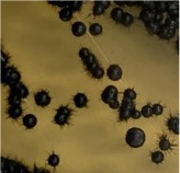
| Fungi, especially several model species like S. cerevisiae (budding yeast),
S. pombe (fission yeast), and A. nidulans (filamentous fungi), have made
significant contributions to the fields of biology and medicine, playing a
crucial role in elucidating fundamental cellular mechanisms. However,
recent research has unveiled a diverse range of fungal species inhabiting
marine environments, many of which exhibit distinct cell growth and
division patterns compared to model fungi. For example, several black
yeast species collected at Sugashima MBL have shown the ability to adapt
their growth and division strategies in response to environmental cues (a
phenomenon known as phenotypic plasticity). We aim to conduct cuttingedge
cell biology research on fungi originating from marine sources. |
 |
|
(Fig. 1)Black yeast collected at Sugashima MBL |
|
|
Marine macroalgae (seaweeds)
|
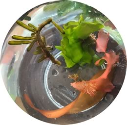
| Sugashima is renowned for its diverse array of seaweeds, including nori
(a seaweed commonly used in Japanese cuisine). Our research delves into
the cellular intricacies of these seaweeds. Many macroalgal species
exhibit cellular characteristics that markedly differ from those of
terrestrial organisms. For example, while cell division is a fundamental
process for all living organisms, seaweeds showcase unconventional
patterns even in this activity, challenging conventional understanding.
When seeking to understand these phenomena, it is not possible to
directly apply insights gleaned from research on model land plants.
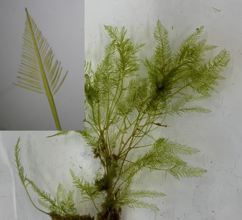
Presently, our focus is centered on the study of the green algae Bryopsis.
This macroalga, which grows to ~10 cm, resembling bird feathers, is
remarkably a single-celled organism containing multiple nuclei (called
coenocyte). We are intrigued by how it forms intricate shapes without
undergoing cell division. |
|
|
(Fig. 2)Macroalgae collected at Sugashima MBL. Right; Bryopsis sp. |
|
|
|
|